Mobile app
Capture fields. Skip the manual tally.
- Smartphone-camera acquisition over the microscope optics
- Deep object-counting model running inside the app
- Thick-smear and thin-smear research workflows
- Local operation when the laboratory is offline
RESEARCH SOFTWARE / PLASMODIUM FINDER
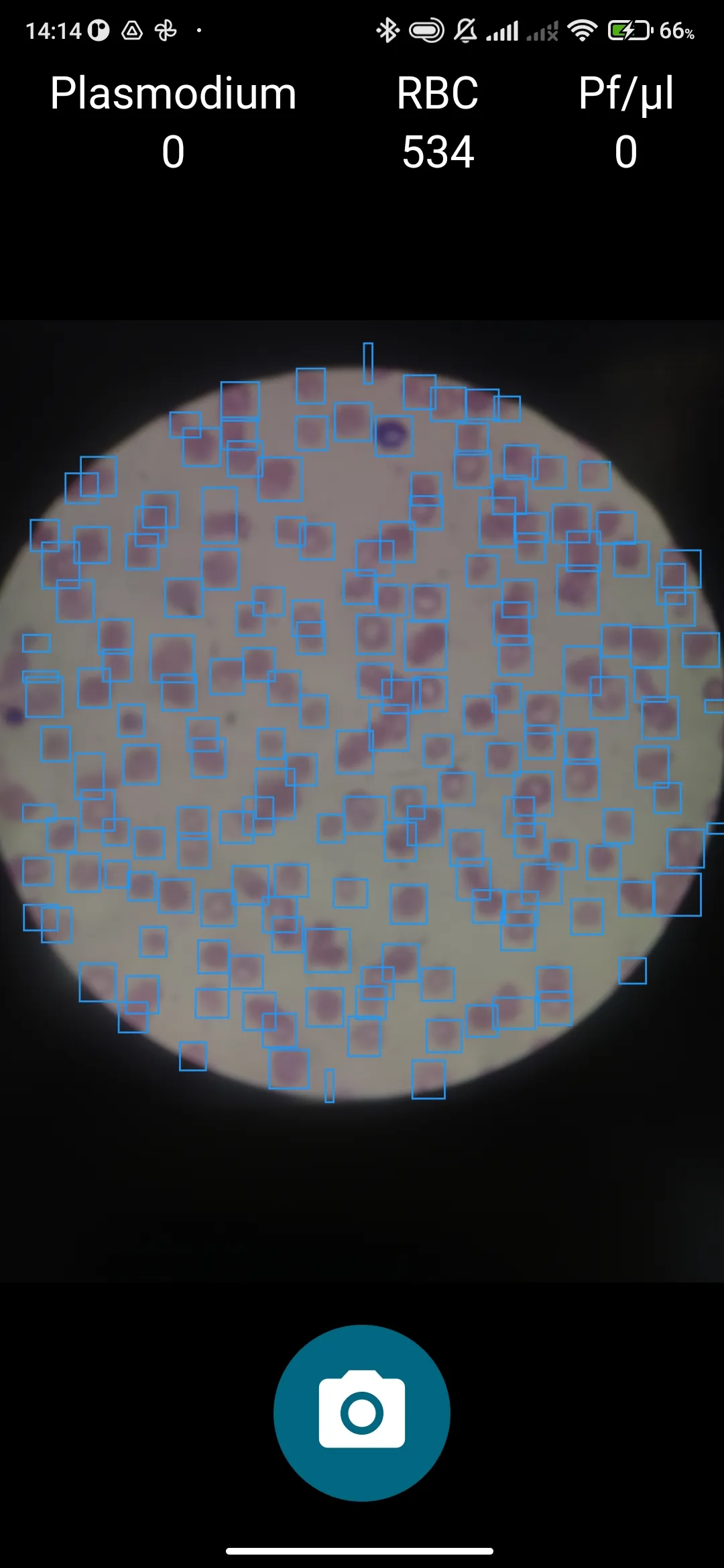
Plasmodium Finder removes the repetitive manual-counting step from ring-stage P. falciparum research. Move across the slide, press capture, and the on-device model quantifies each acquired field.
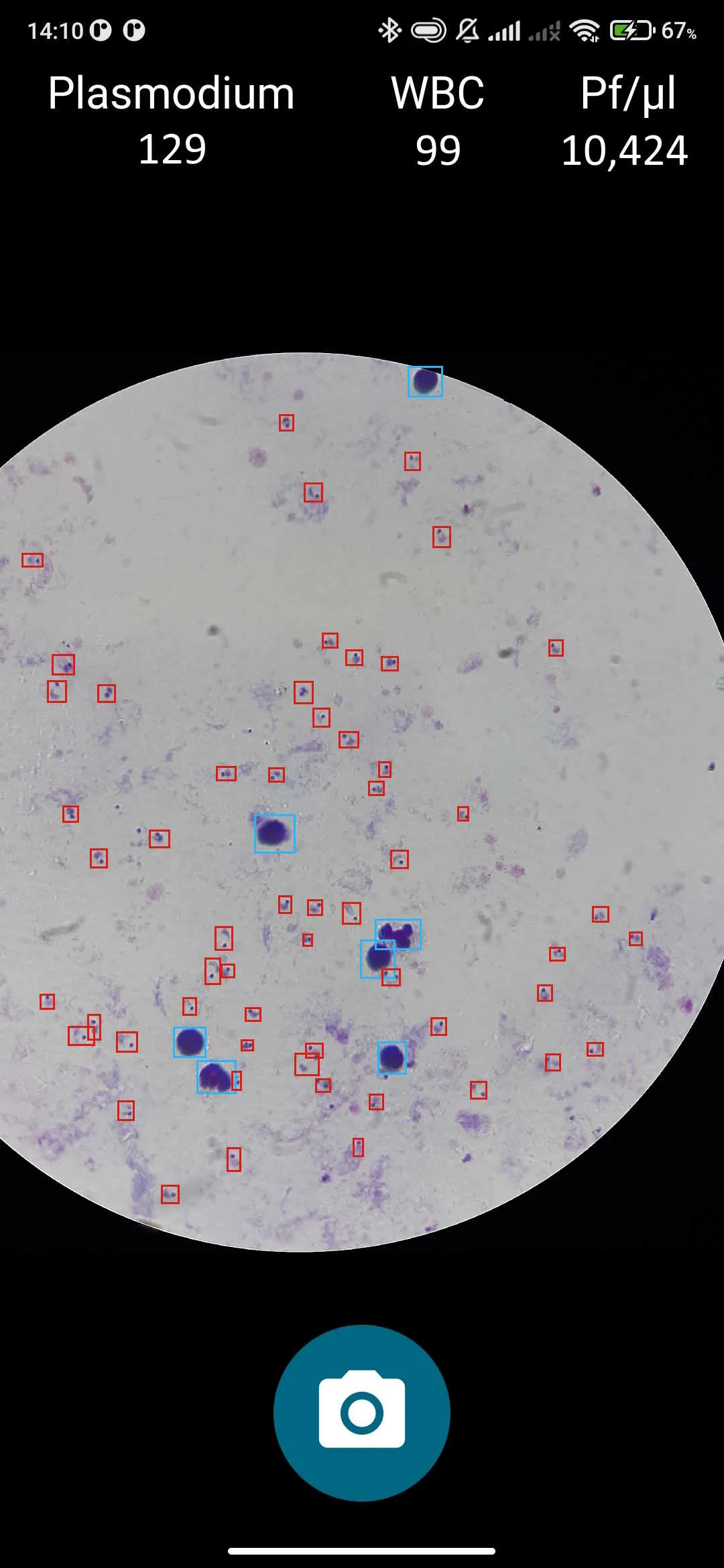
Position the smartphone camera over the microscope optics and move through the slide as usual. Each press of the capture button adds a field to the acquisition, while the model handles the ring-stage object count inside the app.
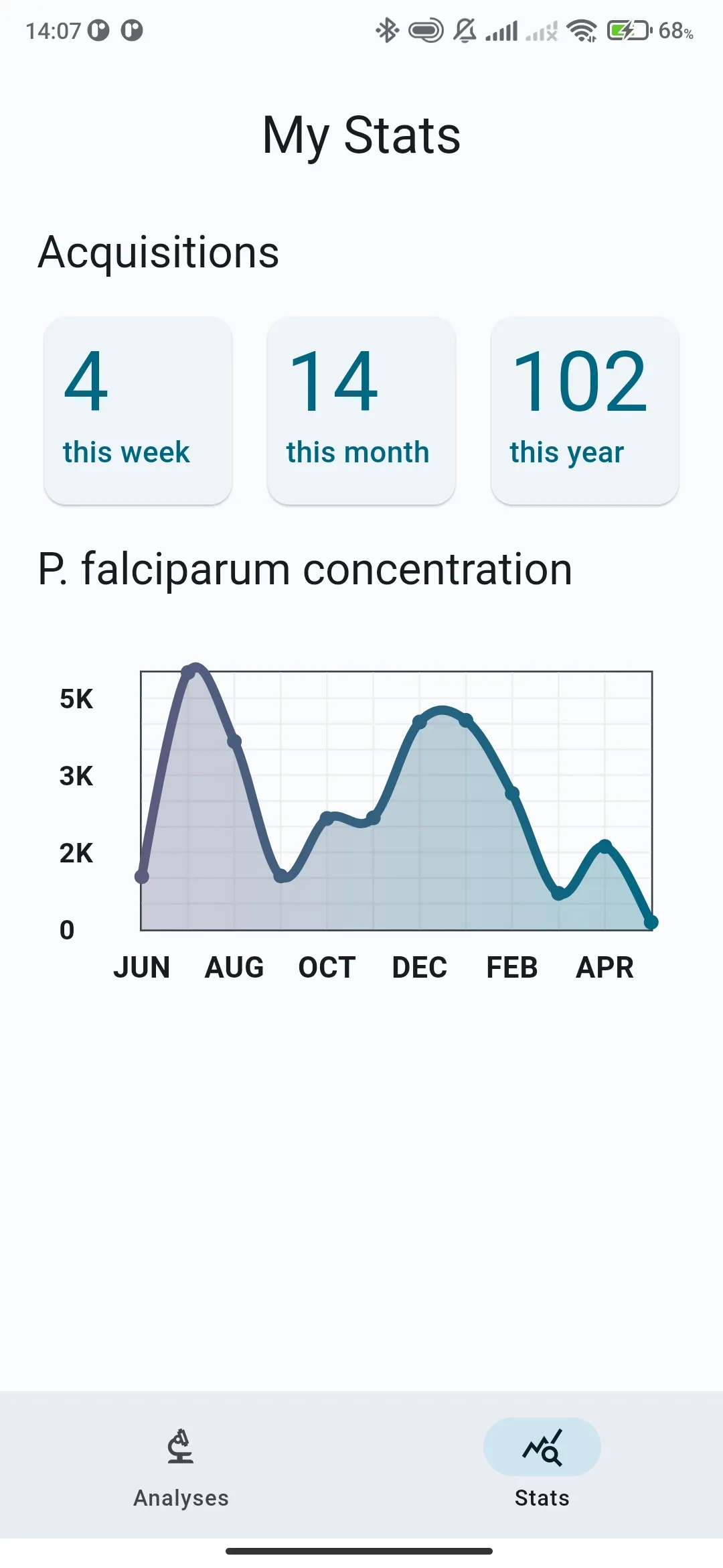
The mobile app handles acquisition and counting. When a connection is available, the web workspace makes laboratory results available to view and export.
Mobile app
Web workspace
Position a compatible smartphone above the microscope optics using an appropriate adapter.
Capture thick- or thin-smear fields from the mobile application.
Let the on-device model quantify ring-stage objects instead of tallying each captured field by hand.
Upload results when online, then view and export them from the web workspace.
Pair the software with a Micrensa microscope adapter to position the smartphone camera over the optics for repeat acquisition work.
Tell us about your research use, smear types and acquisition workflow to discuss access for your laboratory.